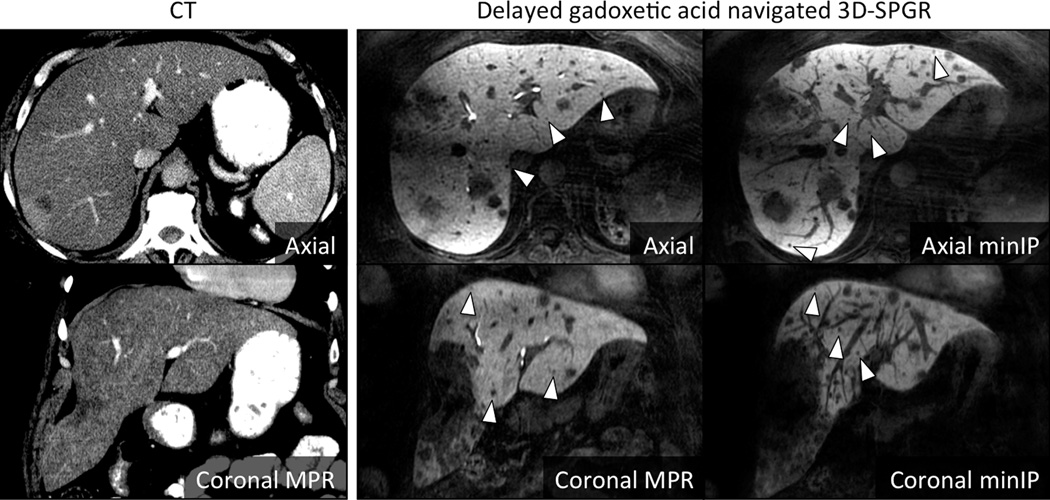
Figure 5. Visualization of non-hepatocyte origin intrahepatic neoplasm.
In this patient with colorectal adenocarcinoma metastases, the high conspicuity of the low signal lesions against the high signal liver is apparent. Note the small size of the lesions (arrowheads) made more apparent on the minimum intensity projection images (right panels), many of which were undetectable on CT (left panels).

