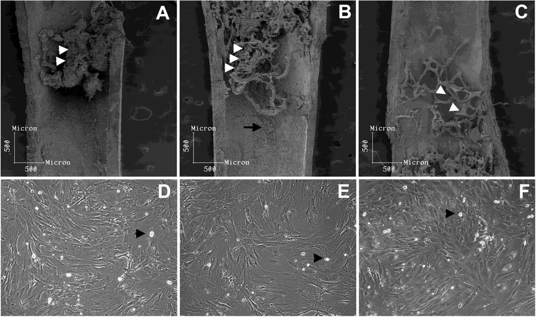
Figure 1. Analysis of the inner surface of femurs and morphology of the stromal populations isolated.
After bone marrow was flushed, subendosteal and endosteal stromal cells which remained attached were mainly distributed in the trabecular region (A, white arrow heads) at the metaphysis and epiphysis. After the first round of collagenase digestion procedure, subendostal stromal cells were removed (B) and osteoblasts remained attached (B, white arrow heads and arrow), being retrieved only after the second collagenase digestion (C, white arrow heads pointing to osteoblast-free trabecular bone). When in culture, flushed bone marrow (D) and subendosteal stromal cells (E) presented similar myofibroblastic morphology. Conversely, endosteal osteoblats (F) presented a cuboidal morphology, as expected. A few macrophage-like cells were observed in all three primary cultures (arrow heads). Primary cells were cultured to confluence in DMEM 10% FBS.

