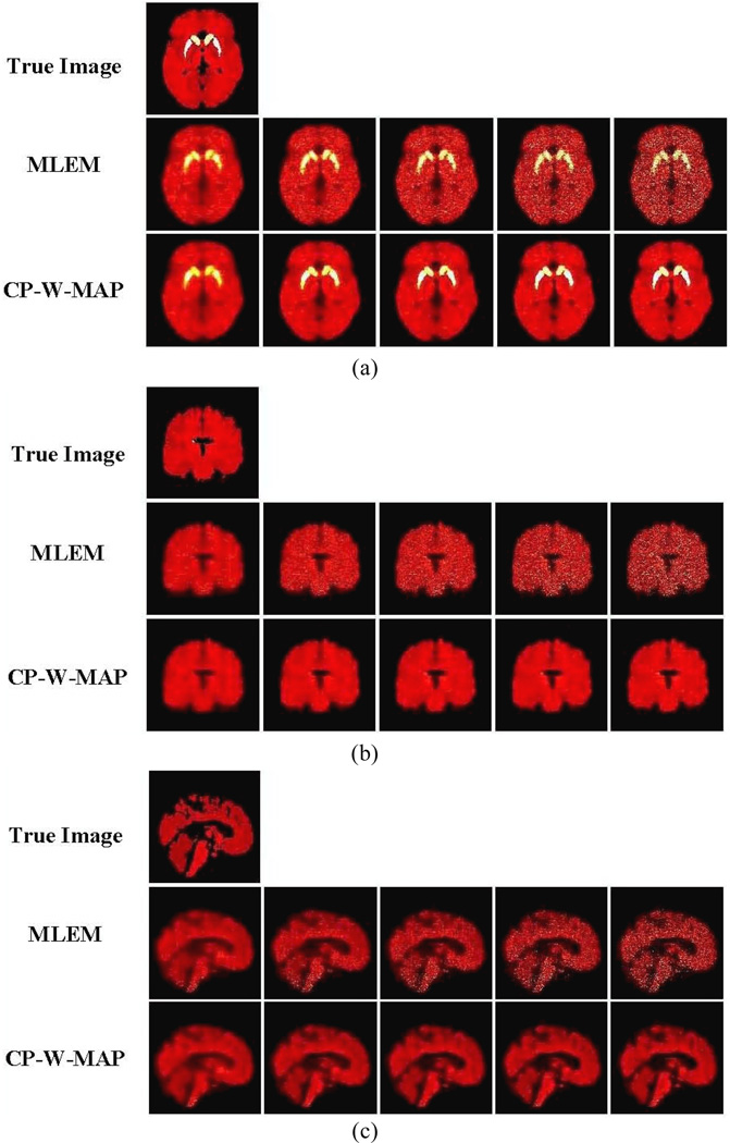
Figure 8.
True and estimated parametric DV images: (a)–(c) corresponding to transaxial, coronal and sagittal slices, respectively. For each, (i) true image, (ii) standared 3D MLEM reconstruction (MLEM) and (iii) proposed 3.5D reconstruction (CP-W-MAP) are shown (From left to right): Increasing iterations of 1, 2, 3, 5 and 10 (16 subsets). No post-filtering was applied to the images shown.

