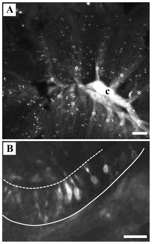
Figure 4.
DiI labeling of OSNs from lateral olfactory bulb. A) When the lipophilic tracer DiI was inserted into the lateral region of the olfactory bulb, the retrogradely labeled axons coalesced in the central raphe (c). Labeled profiles representing olfactory sensory neurons were seen randomly distributed throughout the sensory region of the olfactory rosette along the lamellae (*). Scale bar=100 μm. B) Higher magnification analysis of DiI-labeled rosettes allowed examination of the morphology of the olfactory sensory neurons that project to the lateral olfactory bulb. The majority of OSNs observed in the olfactory epithelium by retrograde labeling had cell bodies in the middle of the epithelium and intermediate-length dendrites. Dashed line=apical surface, solid line=basement membrane. Scale bar=20 μm.

