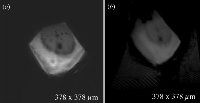
Figure 11.
On-axis image of an X-ray-exposed lysozyme crystal compared with an ‘on-axis-like’ view of the same crystal reconstructed from an off-axis microscope direction. Confocal microscopy requires only one camera direction to reconstruct a full volumetric model of the sample. Off-axis views can be obtained as projections of the volume, but with typically lower resolution that is determined by the number of focal planes scanned and by the point-spread function normal to those planes. While the on-axis view (a) gives the maximum resolution in this experiment, the same view reconstructed from a confocal scan taken 90° off axis (b) still resolves important features of the crystal.

