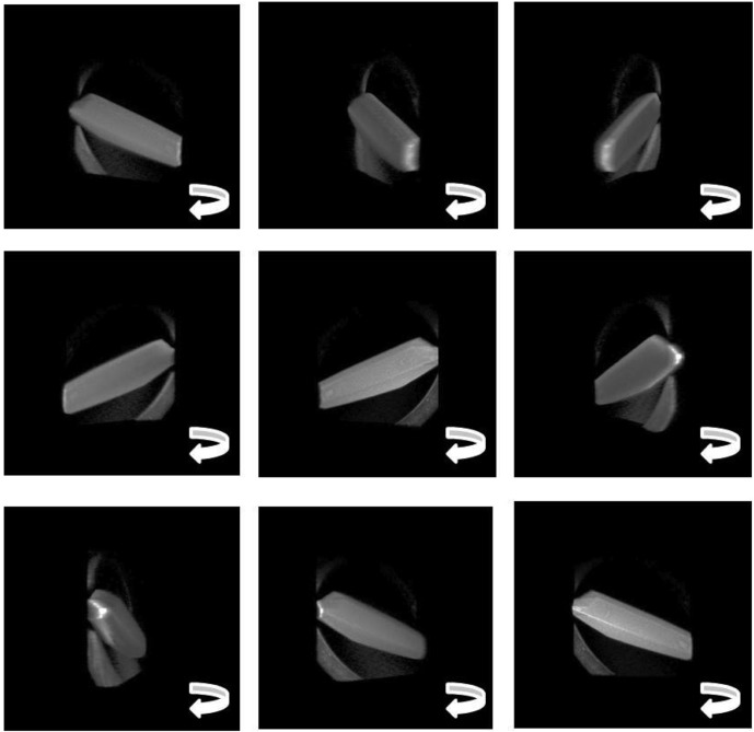
Figure 5.
Stills taken from the reconstructed three-dimensional view of a thermolysin crystal on a fiber mount (supplementary material, movie 3). The three-dimensional view was reconstructed by combining stacks of confocal images (307 × 307 µm) taken in fluorescence mode after the crystal was soaked in acridine orange dye. This series of stacks is shown in Fig. 4 ▶.

