Fig 1.
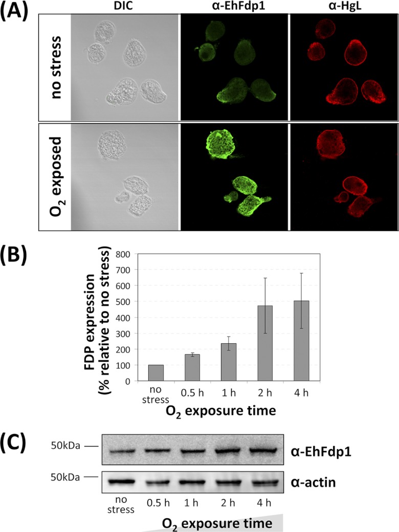
EhFdp1 localizes to the cytoplasm and protein abundance increases upon oxygen exposure. (A) Immunofluorescence microscopy analysis of EhFdp1. Phase-contrast (differential interference contrast [DIC]) transmission images are shown. Antibodies: anti-EhFdp1 (1:500; Open Biosystems), anti-rabbit–Alexa 488 (1:1,000; Molecular Probes), anti-HgL (1:50), and anti-mouse–Alexa 594 (1:500; Molecular Probes). Images were collected with a Zeiss LSM700 confocal microscope and filtered using Volocity software (Improvision). (B) Western blot analysis of the effect of oxygen exposure on EhFdp1 expression in E. histolytica HM-1:IMSS. Anti-EhFdp1 antibody (1:500) and HRP-conjugated anti-rabbit antibody (1:1,000; Cell Signaling) were used (1 h of incubation each), and the signal was detected with ECL+ (GE Healthcare). Primary (anti-actin, 1:1,000; MP Biomedicals) and secondary (HRP-conjugated anti-mouse antibody at 1:1,000; Cell Signaling) antibodies were used for the actin control. Densitometric analysis with ImageJ reveals an increase in EhFdp1 expression with increasing oxygen exposure time from ∼2-fold after 1 h (n = 5; P = 7.0 × 10⁻³) to ∼5-fold after 4 h (n = 5; P =: 0.029). (C) Representative Western blot assay displaying EhFdp1 levels increasing with oxygen exposure time.

