
Fig 1.
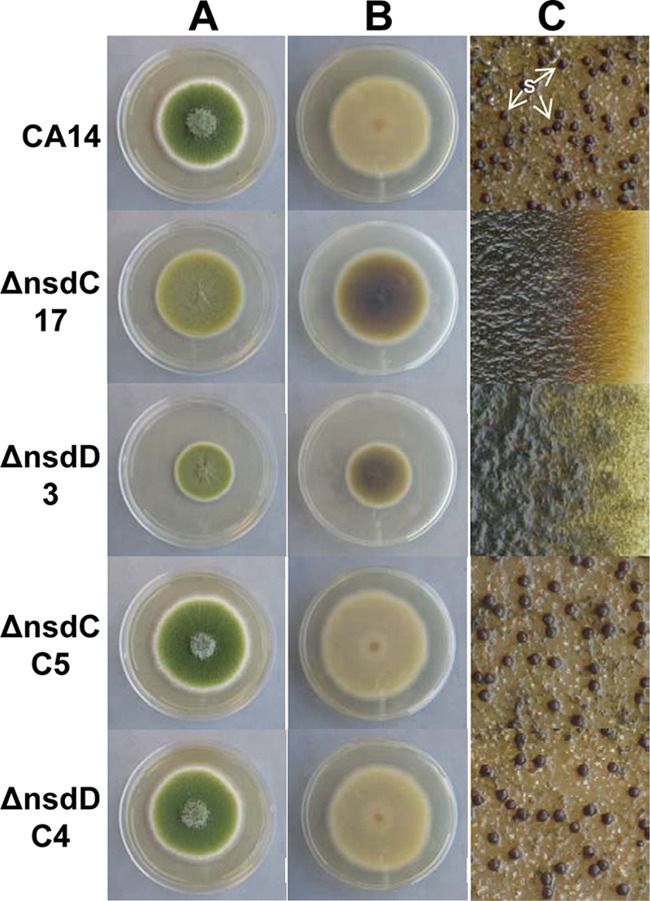
Colony growth, pigmentation, and sclerotium production in A. flavus CA14 ΔnsdC and ΔnsdD mutants and ΔnsdC and ΔnsdD complementation strains. CA14, ΔnsdC 17 and ΔnsdD 3 mutants, and genetically complemented ΔnsdC C5 and ΔnsdD C4 strains were grown on YGT-U medium for 5 days with fluorescent light illumination. (A) View from top of colony. Conidial pigmentation was altered in the Δnsd mutants. Decreased growth of the ΔnsdD 3 strain was observed. (B) View of underside of colony. Pigmentation was readily observed on the undersides of the ΔnsdC 17 and ΔnsdD 3 mutant colonies. (C) YGT-U plates demonstrating sclerotium production were grown for 14 days in the dark. The plates were sprayed with 70% ethanol to allow visualization of sclerotia. Sclerotia were absent in the ΔnsdC and ΔnsdD mutants and were produced in the wild-type CA14 and ΔnsdC and ΔnsdD complementation strains. S, sclerotia.