Figure 3.
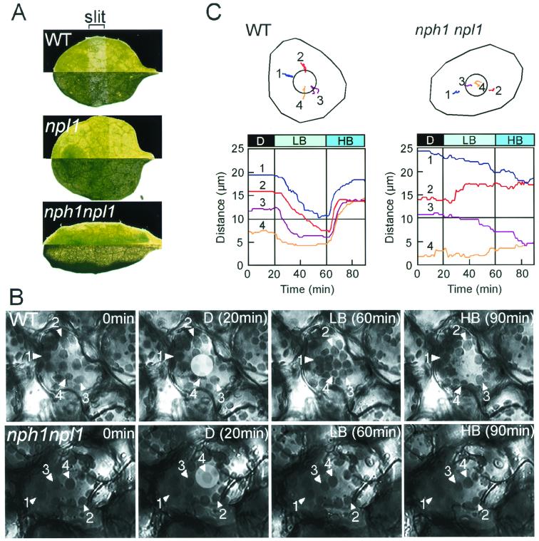
Light-activated chloroplast relocation in wild-type plants and the nph1npl1 double mutant. (A) Slit assay for chloroplast relocation. Leaves from wild-type (Columbia), npl1-101, and the nph1npl1 double mutant were partially irradiated with high-intensity blue light for 1 hr. Photographs taken in dark (upper section) and bright (lower section) fields of vision are shown in each case. (B) A series of images monitoring chloroplast relocation in single mesophyll cells from wild-type (Columbia) plants and the nph1npl1 double mutant. Chloroplast accumulation movement was induced by continuous microbeam irradiation with low-intensity blue light (LB, 2 μmol⋅m−2⋅s−1) from the 20th to 60th minute after the onset of the experiment (D, without blue light irradiation). Chloroplast avoidance movement was induced with high-intensity blue light (HB, μmol⋅m−2⋅s−1) from the 60th to the 90th min. The microbeam (20 μm in diameter) used can be seen as a light circle in each image taken after 20 min. (C) Movement tracks of individual chloroplasts. The movement of each chloroplast numbered in B was traced during the experiment and is shown in Upper. Circles in the cells represent the irradiated areas. Distances (μm) between the beam center and each chloroplast were also recorded and are shown in Lower. The 10-μm distance represents the range of the irradiated area (the radius of the blue light microbeam).

