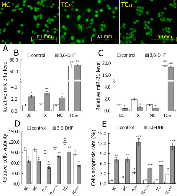
Figure 5.
Increasing intracellular miR-34a or silencing miR-21 expression promoted 3,6-dihydroxyflavone-induced breast cancer cell cytotoxicity and apoptosis. (A) Photomicrographs of transfected cells by fluorescence microscopy. (B) Expression of microRNA-34a (miR-34a) in each cell and the effect of 3,6-dihydroxyflavone (3,6-DHF). (C) Expression of microRNA-21 (miR-21) in each cell and the effect of 3,6-DHF. (D) Effect of 3,6-DHF (10 μM) treatment for 24 hours on cell viability. (E) Effect of 3,6-DHF (10 μM) treatment for 24 hours on cell apoptosis. Data presented as mean ± standard deviation (n = 3). *P < 0.05, **P < 0.01 compared with control in each group. #P < 0.05, ##P < 0.01 compared with the MDA-MB-453 breast cancer cell (BC) group. TX, tumor xenograft of MDA-MB-231 breast cancer cells; MC, MDA-MB-453 cells transfected with blank plasmids (mock cells); TC34a, MDA-MB-453 cells transfected with pcDNA6.2-GW/miR-34a; TC21, MDA-MB-453 cells transfected with pcDNA6.2-GW/miR-21. All transfected cells were collected for the next experiments 48 hours after transfection.

