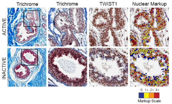
Figure 2.
Active phenotype is associated with increased density of TWIST positive cells. Representative Active and Inactive tissues are shown. Two trichrome images are shown (different magnifications) and the TWIST1-DAB stained section illustrates that despite a greater density of epithelial cells in the Inactive patient, there is a lower density of TWIST staining and the intensity of staining (nuclear markup image) is reduced relative to Active patients. The nuclear markup scale ranges from blue (no staining) to red (3+ intensity).

