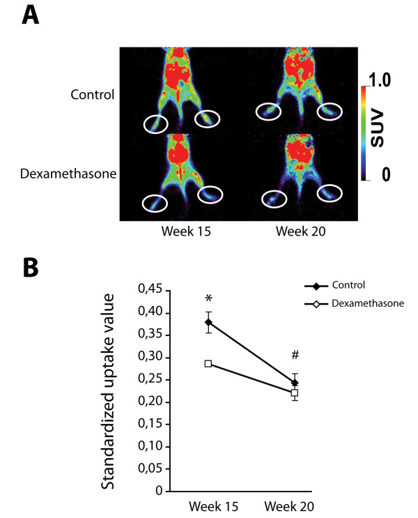
Figure 3.

Dexamethasone treatment influences the inflammatory component of spontaneous arthritis as measured by 18F-FDG PET scan. (A) Representative images of animals from both treatment groups. The 'toe' area used for measurements is indicated. (B) Standardized uptake values of the tracer demonstrating a reduction in the uptake after dexamethasone treatment at week 15. At week 20, the inflammatory component has largely disappeared as seen in the control group. (n = 4 animals in dexamethasone treated group and 6 control animals; * P < 0.05 dexamethasone treatment versus control group; # P < 0.05 week 20 versus week 15 in control group). 18F-FDG, 2-[18F]fluoro-2-deoxy-D-glucose; PET, positron emission tomography.
