
Figure 1.
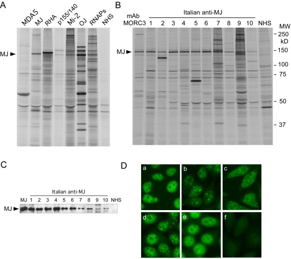
Detection of anti-MJ antibodies. A. Immunoprecipitation of anti-MJ and other autoantibodies that recognize proteins of similar molecular weight. 35S-methionine labeled K562 cell extract was immunoprecipitated by human sera and analyzed by 8% SDS-PAGE. Anti-MJ serum immunoprecipitates a ~140 kDa protein (arrow), which is different from the mobility of other known autoantigens. Reference sera for anti-MDA5, -MJ, -RNA helicase A (RHA), -p155/140, -Mi-2, OJ, and -RNA polymerases (RNAPs) are shown. NHS, normal human serum. B. Immunoprecipitation of anti-MJ positive sera. 35S-methionine labeled K562 cell extract was immunoprecipitated using mouse anti-MORC3 monoclonal antibody (lane mAb MORC3), human anti-MJ positive sera (lanes 1 to 10) or a normal human serum (NHS), and analyzed by 8% SDS-PAGE. MW, molecular weight marker. C. IP-Western blot of MJ. The identity of the 140 kDa protein as MJ/NXP-2/MORC3 was verified by IP-Western. The MJ protein is indicated with the arrow. MJ, anti-MJ reference serum, lanes 1 to 10, anti-MJ positive Italian samples. NHS, normal human serum. D. Immunofluorescence staining of anti-MJ positive sera. HEp-2 slides were stained with mouse anti-MORC3 monoclonal antibody (a), human anti-MJ (+) sera (b) to (e), or normal human serum (f). Serum dilution, 1:80.