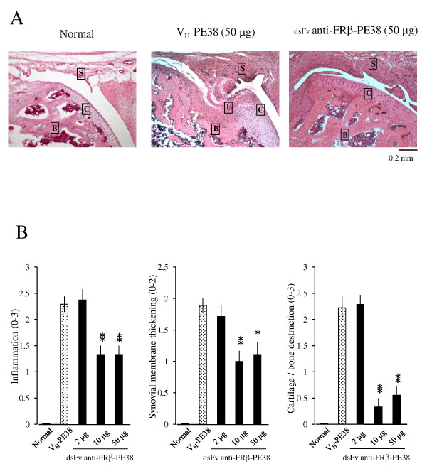
Figure 5.

Effects of an anti-FRβ immunotoxin on the histology of knee joints in rat AIA. (A)Photographs are representative of knee joints from eight rats per group. Knee joints from normal rats and AIA rats treated with 50 µg of VH-PE38 or dsFv anti-FRβ-PE38 were stained using H & E. Note that a knee joint from an AIA rat treated using dsFv anti-FRβ-PE38 showed less cartilage/bone destruction compared to a rat treated using VH-PE38. Letters S, C, B, and E represent synovium, cartilage, bone, and erosion. Original magnification was ×100. (B)Histological scores were recorded for inflammation, synovial membrane thickening, and cartilage/bone destruction. Data are presented as the mean ± SEM of each histological score from eight rats per group. *P < 0.05 and **P < 0.01 compared to the group treated using VH-PE38. AIA, antigen-induced arthritis; FRβ, folate receptor β; H & E, haematoxylin and eosin; SEM, standard error of the mean.
