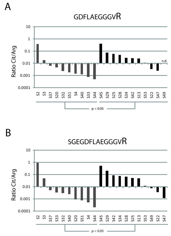
Figure 5.

Relative abundance of citrullinated peptides and their noncitrullinated counterparts. Bar graphs showing the ratio between the abundance of a specific citrullinated peptide versus its noncitrullinated variant in rheumatoid arthritis (RA) patients (black bars) and control patients (gray bars) on a base 10 logarithmic scale. Panel A shows the ratios for peptide GDFLAEGGGVR and panel B for peptide SGEGDFLAEGGGVR. When no signal for the peptide was detected, a value equivalent to the background signal was assigned for this calculation. If both the signal of the citrullinated and the noncitrullinated peptide were missing, the ratio is shown as not determined (n.d.). Patients are sorted per group according to the observed ratios.
