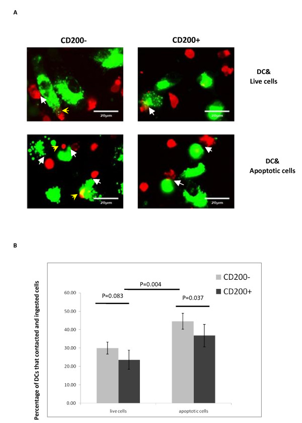
Figure 8.

CD200 expression on apoptotic cells affected their binding and phagocytosis by dendritic cells. (A) Immature dendritic cells (DCs) (stained with PKH67, green) were incubated with target cells pre-labeled with PKH26 (red) and examined under immunofluorescence microscopy. White arrowheads, target cells attached to immature DCs; yellow arrowheads, target cells engulfed by immature DCs. Upper left, CD200- live cells; upper right, CD200+ live cells; lower left, CD200- apoptotic cells; lower right, CD200+ apoptotic cells. (B) Quantification of the percentages of DCs that bound and ingested target cells by immunofluorescence microscopy. The percentages were calculated by counting all the cells in 10 microscope fields (average 40 to 50 cells each microscope field and 400 to 500 cells were recorded in total).
