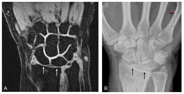
Figure 2.

JSN depiction in wrist joints. Coronal fat-suppressed T1-weighted gradient-echo image of wrist (A) shows sharp delineation of bone margins free of chemical-shift artifacts, thus allowing accurate determination of the joint-space widths, corresponding closely to those seen with XR in the same wrist (B). Note how clearly both MRI and XR show the radius-lunate and radius-scaphoid joint spaces (arrows) to be narrowed (JSN) relative to the other joints in the field of view. A JSN score of 2.0 was given to these joints independently on MRI and XR. MRI (A) additionally depicts the articular cartilage directly as high-signal tissue lining the articular cortices of the bones and showing sharp contrast with adjacent low-signal joint fluid on one side and low-signal articular bone and subcortical marrow fat on the other. As illustrated in this example, the interfaces between opposing cartilage surfaces, particularly in normal joints, often can be sharply delineated. Note that although the radius-lunate and radius-scaphoid joints are clearly narrowed on both MRI and XR, MRI further shows the cartilage to be only thinned on both sides of these joints, without complete denuding in any location. This important distinction cannot be determined with XR.
