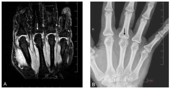
Figure 3.

Joint-space narrowing (JSN) depiction in metacarpophalangeal (MCP) joint. Coronal, fat-suppressed, T1-weighted gradient-echo image of MCP joints 2 through 5 (A), and corresponding XR image (B) show grade-2.0 JSN of MCP 3 (arrow) but normal joint-space width of MCP 2, 4, and 5. The proximal interphalangeal joints are out of the plane of section on the MR image shown, but were visible on more-palmar sections of the scan (not shown).
