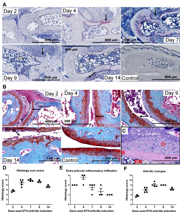
Figure 2.

Development of severe arthritis in mice with delayed-type hypersensitivity (DTH) arthritis. (a) Histochemical stains for the osteoclast-specific enzyme tartrate-resistant acid phosphatase (TRAP). Osteoclasts appear red. Arrows indicate areas with increased osteoclast numbers and bone erosion. (b) Histochemical stain using the Safranin O protocol. Cartilage proteoglycan is stained red, and the intensity of the red color is proportional to the proteoglycan content of cartilage. Arrows indicate areas displaying cartilage loss, which is most prominent in the uncalcified cartilage. Mice with DTH arthritis developed severe arthritis characterized by increased cartilage degradation (as assessed by loss of Safranin O staining) and osteoclast activity (as assessed by the increase in TRAP-positive cells) and bone erosion; synovitis and pannus formation is also observed. The sections shown in (a) and (b) are representative of mice with DTH arthritis (immunization, anti-type II collagen antibody cocktail (anti-CII), and antigen challenge) and of mice receiving immunization, anti-CII, and no challenge (control). Samples were taken on days 2, 4, 7, 9, and 14. (c) Osteophyte shown on day 14 after DTH-arthritis induction. Hematoxylin and eosin staining was used. B, bone; BM, bone marrow; O, osteophyte. (d) Total sum score of histopathological changes from days 2 to 14 after DTH-arthritis induction. Maximum possible score is 15. Mean ± standard error of the mean (SEM) is shown (n = 3 to 5). (e) The score for extra-articular inflammatory infiltration from days 2 to 14 after DTH-arthritis induction. Maximum possible score is 3. Mean ± SEM is shown (n = 3 to 5). (f) Sum score of arthritic changes (calculated as total sum score of histopathological changes from which the score for extra-articular inflammatory infiltration has been subtracted) from days 2 to 14 after DTH-arthritis induction. Maximum possible score is 12. Mean ± SEM is shown (n = 3 to 5). Details of the histopathological scoring system can be found in the Materials and methods section.
