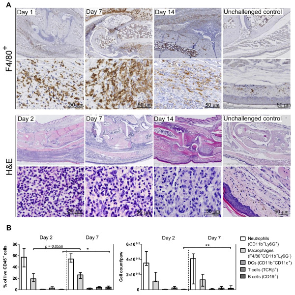
Figure 3.

Composition of the inflammatory infiltrate in delayed-type hypersensitivity (DTH) arthritis. (a) Representative immunohistochemical stains for F4/80+ cells and hematoxylin and eosin (H&E) stains from mice receiving immunization, anti-type II collagen antibody cocktail (anti-CII), and challenge. Samples were taken on days 1 (F4/80+), 2 (H&E), 7, and 14 after DTH-arthritis induction. Unchallenged control samples are from mice receiving immunization, anti-CII, and no challenge. Mice with DTH arthritis displayed severe inflammation characterized by an infiltration of neutrophils and F4/80+ cells into the soft tissue and intra-articular space; neutrophils dominated in the early stages and in the intra-articular space. Severe hyperplasia of the synovial membrane and pannus formation were also observed. (b) Flow cytometric analysis of inflammatory infiltrate isolated from inflamed paws. Cell subsets are displayed both as fraction of total live CD45+ cells and as absolute numbers per paw. Cells were gated on singlets, live cells, and CD45+, and cell subsets were defined as follows: neutrophils: Ly6G+CD11bintermediate-high; macrophages: F4/80+CD11b+Ly6G-; dendritic cells (DCs): CD11b+CD11c+; T cells: TcRβ+; B cells: CD19+. Median ± range is shown (n = 5). *P ≤ 0.05, **P ≤ 0.01. TcRβ+, T-cell receptor-beta-positive.
