
Figure 3.
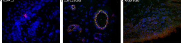
ADORA protein expression. Two-color immunofluorescence detecting ADORA2A (A; red), ADORA2B (B; green) and ADORA3 (C; red) protein expression in combination with cell specific markers (indicated) CD 31(B; red) and CD3 (C; green) in rheumatoid synovia. Co-localized staining appears yellow. ADOR, adenosine receptor.