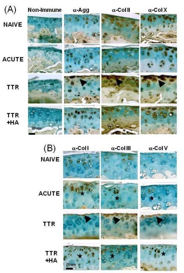
Figure 5.

Immunohistochemical analyses of chondrogenic and fibrogenic matrix molecules in cartilages of naïve and treated mice. Typical examples of immunostained sections for each antibody, and a non-immune isotype control are shown for the four experimental groups (Naïve, Acute, TTR and TTR+HA). Details of antibodies and staining conditions used are described in the Methods Section. (A) Enhanced staining for chondrogenic proteins (A) and fibrogenic proteins (B) in Acute and TTR+HA conditions are highlighted by black stars, and staining in lesional area of the TTR condition are marked by black arrowheads.
