
Figure 6.
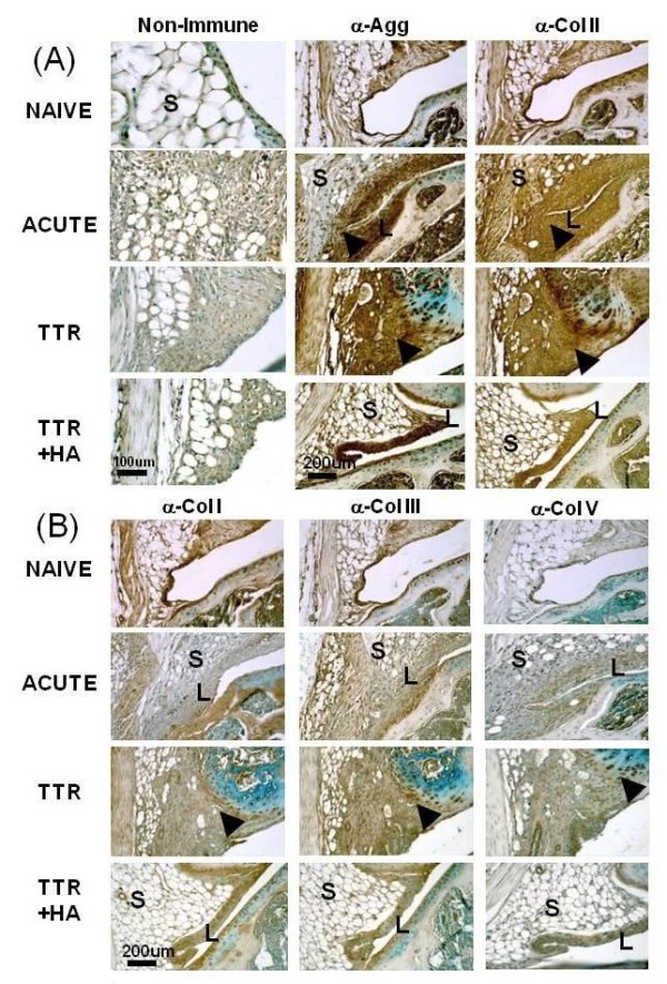
Immunohistochemical analyses of matrix components in the lateral perimeniscal synovium of naïve and treated mice. Typical examples of immunostained sections for each antibody, and a non-immune isotype control are shown for the four experimental groups (Naïve, Acute, TTR and TTR+HA). Enhanced staining of aggrecan and collagen II in the hyperplastic (Acute and TTR+HA) and fibrotic regions (TTR) are indicated by black arrow heads. For the TTR condition, strong staining with anti-collagen II or V was seen in the fibrotic regions, as well as around groups of meniscal fibrochondrocytes, and vascular elements stained positively with anti-collagen V. In all treatment groups, collagen X staining patterns were essentially identical to those seen for collagen II (data not shown). L, Lining; S, Stroma.