
Figure 7.
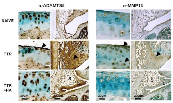
Immunohistochemical analyses of ADAMTS5 and MMP13 in cartilages and perimeniscal synovium of naïve and treated mice. (A) Immunohistochemical staining patterns form ADAMTS5 and MMP13. Chondrocytes throughout the depth of cartilage showed intense cell-associated staining for ADAMTS5 in all treatment groups. Cells and matrix in hyperplastic (TTR and TTR+HA), or fibrotic (TTR) regions of the synovium and cartilage lesions (TTR) also showed abundant immunoreactive ADAMTS5. (B) MMP13 staining was seen only in the pericellular space of chondrocytes in TTR samples, and the matrix at the meniscal/synovial interphase (marked by black arrows).