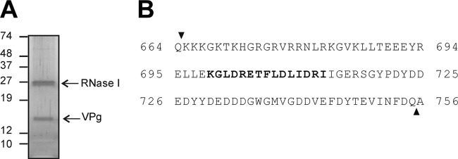
Fig 2.
Silver staining of HAstV VPg and protein identification by mass spectrometry. (A) Total RNA (and proteins associated with it) isolated from cultures of HAstV-4-infected CaCo2 cells at 24 h postinfection was analyzed by SDS-PAGE and silver staining after being treated with RNase I. (B) The 13- to 15-kDa protein band was excised and treated with trypsin for protein identification by Cap-LC-nano-ESI-Q-TOF. The 15-mer peptide identified is indicated in bold over the full length of the predicted HAstV VPg amino acid sequence.

