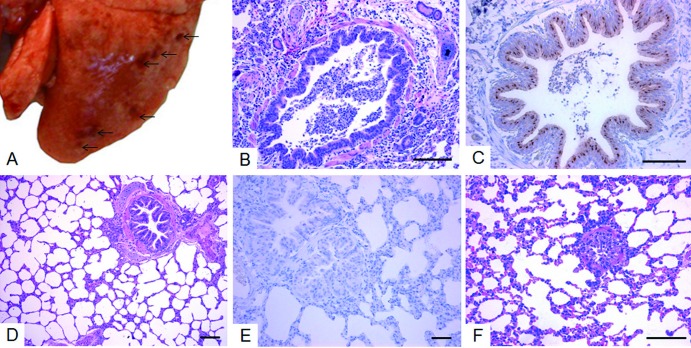
Fig 2.
Lung lesions of ferrets caused by the SC/09 and HA/226R viruses. (A) In the SC/09 virus-inoculated ferrets, macroscopic lesions in the lungs and focal discolorations (arrowed) were observed in all of the lobes. (B) Histologically, the consolidated area in the SC/09 virus-infected ferrets was consistent with the prominent features of bronchointerstitial pneumonia with massive recruitment of lymphocytes into the lumen and surrounding alveoli, sloughing of respiratory epithelium in the peribronchiolar areas, and submucosal edema of the bronchiolar wall (H&E staining; scale bar, 100 μm). (C) Viral antigen was detected in the bronchiolar epithelial cells (IHC; scale bar, 100 μm). (D and E) However, in HA/226R virus-inoculated animals, no obvious histopathological changes were seen in the lung tissues (D) (H&E staining; scale bar, 100 μm) compared to the PBS-inoculated animals (E) (H&E staining; scale bar, 100 μm). (E) Viral antigens were not detected (IHC; scale bar, 100 μm).

