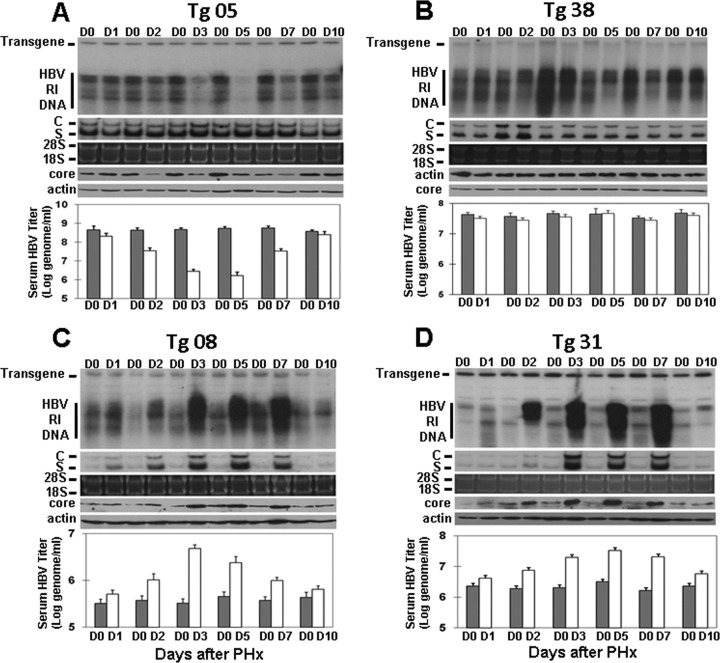
Fig 1.
Effect of a PHx on HBV replication in transgenic mice. Nine-week-old, serum HBeAg-matched male mice were subjected to a 70% PHx and sacrificed at the time points indicated. The liver resected from an individual mouse served as the day 0 (D0) control of that particular mouse. HBV RI DNA, RNAs, and the core protein in the mouse liver were then analyzed by a Southern blot (top panel), a Northern blot (second panel from the top), and a Western blot (fourth panel from the top), respectively. The HBV transgene in the mouse genome was used as the loading control for the Southern blot analysis. 28S and 18S rRNAs were used as the loading controls for the Northern blot analysis (third panel from the top), and α-actin was used as the control for the Western blot (bottom panel). The HBV titers in the serum before (gray bars) and after (white bars) the PHx were measured by real-time PCR and are shown in the histograms. (A) Tg05 mouse line; (B) Tg38 mouse line; (C) Tg08 mouse line; (D) Tg31 mouse line.

