
Fig 6.
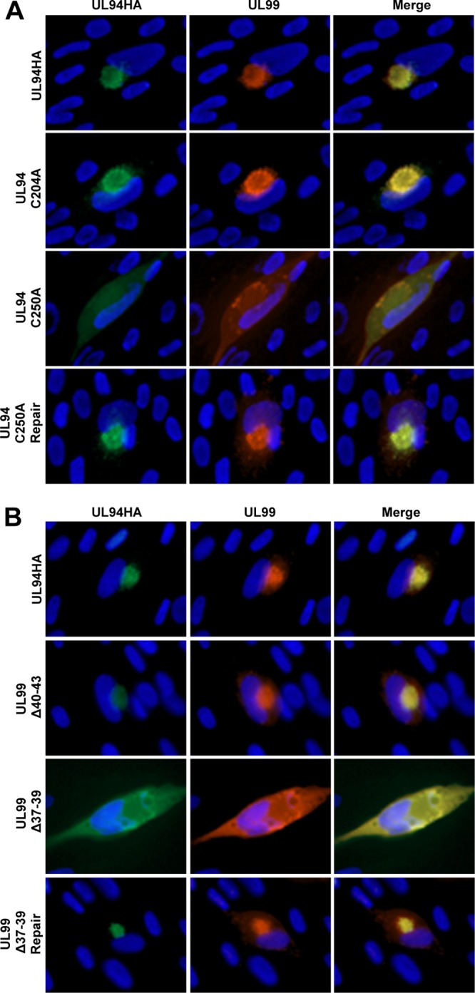
Localization of UL94 and UL99 during infection. (A) HFF cells were infected with the indicated virus at a multiplicity of 0.01 PFU/cell. Cells were fixed at 96 h postinfection, and immunofluorescence analysis was performed with the indicated antibodies to visualize viral proteins. (B) HFF cells were transfected with the indicated BAC DNA. Cells were fixed at 8 days posttransfection, and immunofluorescence analysis was performed with the indicated antibodies to visualize viral proteins. UL94 was detected with anti-HA antibody (green). UL99 was detected with anti-UL99 antibody (red). Nuclei are stained with Hoechst (blue). Images are representative of results obtained from three independent infections (A) or BAC transfections (B).