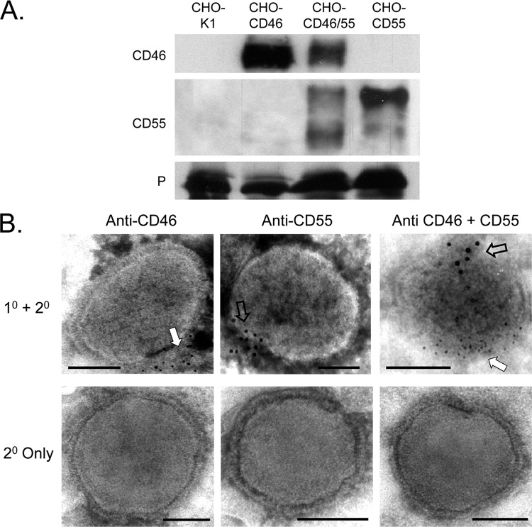
Fig 3.
CD46 and CD55 are associated with purified MuV virions. (A) Western blotting. MuV derived from the indicated cell lines was purified by gradient centrifugation and analyzed by Western blotting for the presence of CD46 or CD55. Levels of viral P protein were analyzed as a loading control. (B) EM analysis. Purified virus derived from CHO cells expressing CD46 (left), CD55 (middle), or both CD46 and CD55 (right) was treated with the indicated antibodies, followed by 6-nm (CD46) or 12-nm (CD55) colloidal gold-labeled goat anti-mouse antibody (top row). Control samples were treated with secondary antibody alone (bottom row). Samples were analyzed by EM at a magnification of ×55,000. Bars, 0.1 μm. White and open arrows, locations of staining for CD46 and CD55, respectively; 1° and 2°, primary and secondary antibodies, respectively.

