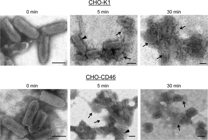
Fig 9.
Electron micrographs of in vitro neutralization of VSV derived from CHO-K1 and CHO-CD46 cells. Purified VSV particles derived from the indicated CHO cells were incubated for the indicated times at 37°C with NHS and then analyzed by negative staining and EM. Arrowheads, apparently intact virions; arrows, clearly lysed particles or nucleocapsid-like structures. Bars, 0.1 μm.

