
Fig 3.
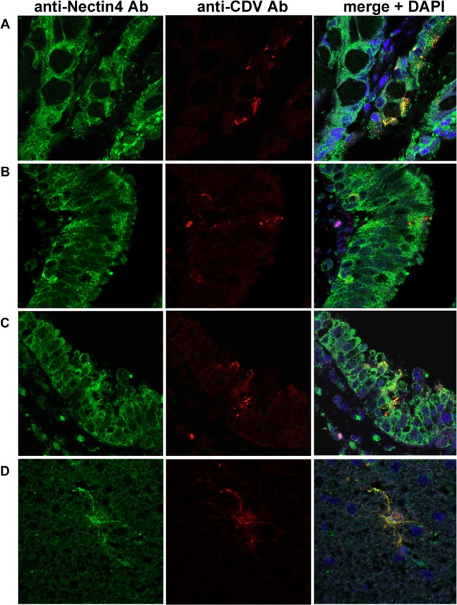
Immunofluorescence double staining for CDV antigen and nectin4. Tissue sections of 2-μm thickness were deparaffinized, rehydrated, and subjected to heat-induced antigen retrieval before incubation with antibodies. CDV antigen was stained with a mouse anti-CDV monoclonal antibody (Adtec, Japan) and Alexa Fluor 594-conjugated secondary (red) antibody. Nectin4 was stained with a goat anti-nectin4 polyclonal antibody (R&D Systems) and Alexa Fluor 488-conjugated secondary antibody (green). Nuclei were counterstained with 4′,6-diamidino-2-phenylindole (DAPI; blue). (A) Intestine; (B and C) lung; (D) brain.