
Figure 1.
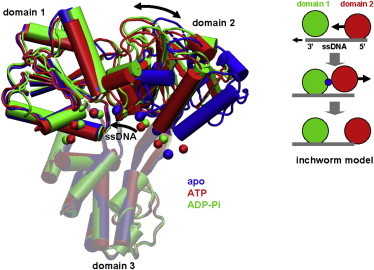
Structures of NS3hel (cartoon representation) bound with ssDNA (chain of beads located at C4¢ atoms) in three biochemical states (blue, apo; red, ATP; and green, ADP-Pi). The three domains of NS3hel and ssDNA are labeled. The opening/closing motions of domain 2 and the 5¢-3¢ sliding of ssDNA are marked by arrows. The three structures are aligned along domain 3 (transparent). A schematic cartoon in the inset illustrates the inchworm model (domain 1, domain 2, ATP, and ssDNA are colored green, red, blue, and gray, respectively; the opening/closing motions of domain 2 and the sliding of ssDNA are marked by arrows; domain 3 is not shown).