
Figure 1.
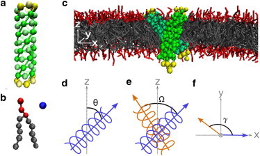
Coarse-grained model and angle definitions. (a) CG model of an α-helix assembled from hydrophobic beads at the core and hydrophilic beads at both ends to keep the helix transmembrane. Helical geometry is maintained by harmonic springs, angle springs, and dihedral angle potentials of principal helix beads (see the Supporting Material for details). (b) CG lipid model includes three hydrophilic headgroups and two five-bead hydrophobic tails. Water is represented explicitly by a single bead. All beads in the system are of the same size and correspond to ∼3 carbon atoms/water molecules. (c) Two positive mismatched helices in a typical crossed configuration (note that, for clarity, water particles are not displayed). (d) Tilt angle, θ, of a helix is defined as the angle between the helix major axis (blue arrow) and the bilayer normal, . (e) Cross angle, Ω, is defined as the dihedral angle between the major axes of the two helices (blue and orange arrows). (f) Projection angle, γ, is defined as the angle between the major helix axes along the plain of the bilayer .