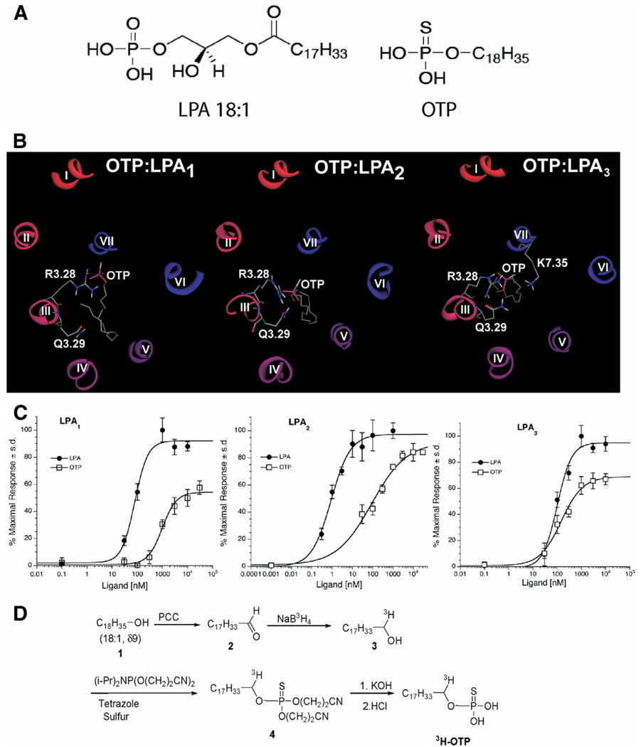
Figure 1.
(A) Chemical structures of LPA and OTP. (B) Molecular models of OTP docked into the ligand-binding pocket of LPA receptors. (C) Ca2+ transients elicited by OTP and LPA in RH7777 cells stably expressing the individual EDG family LPA receptors. Wild-type RH7777 cells show no Ca2+ transients in response to LPA up to concentrations as high as 30 µmol/L (data not shown). (D) Synthesis of 3H-labeled OTP (see Materials and Methods for details).

