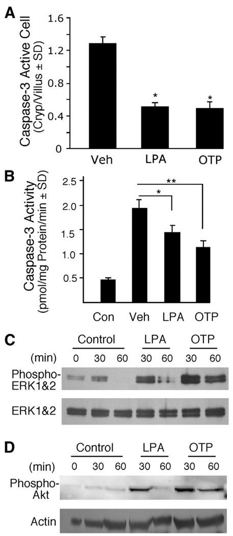
Figure 6.
OTP and LPA reduced caspase-3 activation and activated prosurvival pathways in vivo. C57BL/6 mice were pretreated with 2 mg/kg LPA or OTP for 2 hours and subjected to 15 Gy radiation exposure. Mice were killed 4 hours after radiation. (A) Quantification of active caspase-3 immunoreactive cells. Paraffin-embedded jejunum sections from animals treated with radiation and pretreated with either LPA or OTP were stained with a rabbit polyclonal active caspase-3 antibody and fluorescein-labeled secondary antibody using indirect immunofluorescence as described in Materials and Methods, and the sections were counterstained with 4′,6-diamidino-2-phenylindole (DAPI). Active caspase-3 immunoreactive cells were counted in a minimum of 100 crypt-villus units in groups of 4 animals in the groups. Both agents significantly reduced the number of activated caspase-3–positive cells (*P< .05) when compared with vehicle-treated controls. (B) Caspase-3 activity was determined in epithelial cell lysates prepared from the same animals whose jejunum segments were used for activated caspase-3 immunostaining in A. LPA and OTP both significantly reduced caspase-3 activity in the tissue lysates, a finding in agreement with the reduced number of active caspase-3 cells. (*P< .05, **P< .01) (C and D) Jejunum tissue from mice orally treated with vehicle, 2 mg/kg LPA, or OTP for the indicated times was homogenized and lysates were analyzed using Western blotting with (C) anti–phospho-ERK1/2 or (D) anti–phospho-AKT antibodies and appropriate antibodies to the non-phosphorylated forms of the kinases to monitor equal loading. Note that the activation of both kinases was more robust and longer lasting for OTP compared with LPA.

