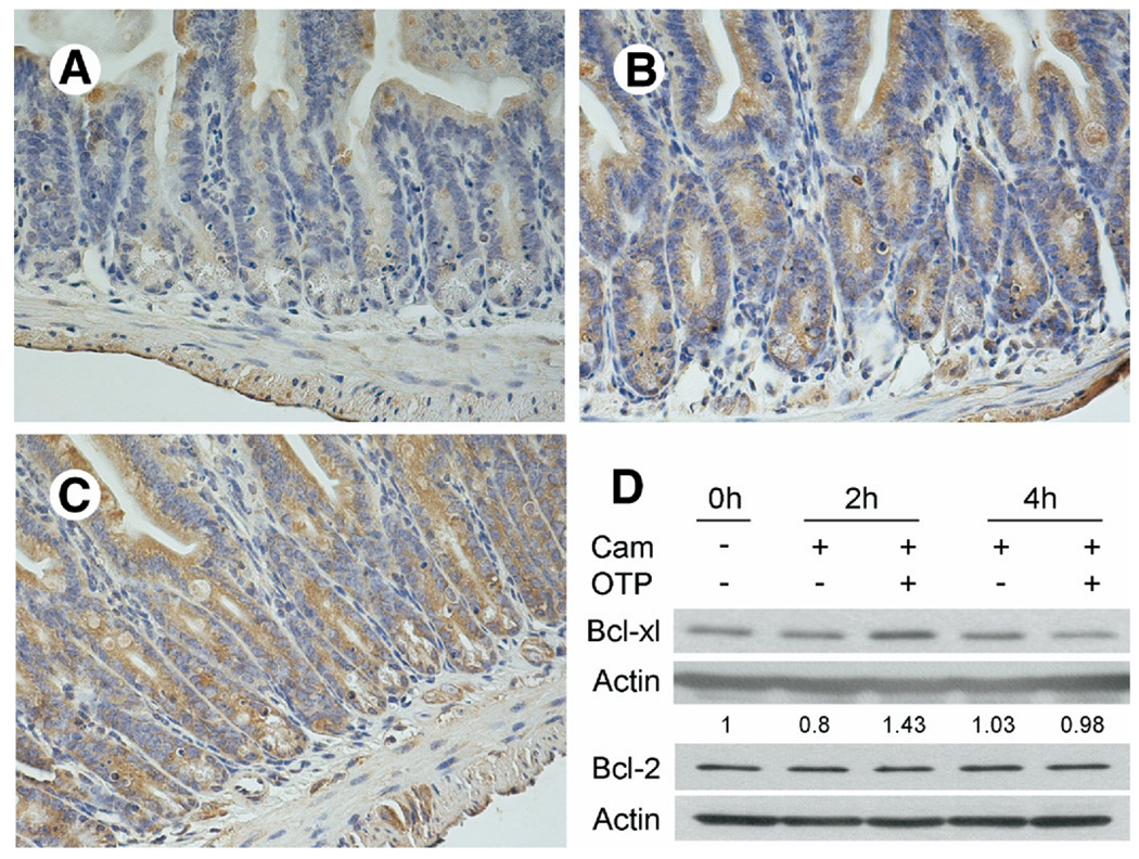
Figure 7.
Immunohistochemical localization of antiapoptotic Bcl-XL protein in vehicle- (200 µmol/L BSA), LPA-, and OTP-pretreated (2 mg/kg each) irradiated small intestinal sections of C57BL/6 mice. Sections of (A) vehicle-treated mice showed less Bcl-XL expression compared with sections obtained from mice treated with (B) LPA or (C) OTP. Calibration bar = 200 µm. The patterns shown are representative to sections obtained from all mice in the corresponding group of 4 mice per treatment. (D) OTP increases the cytoplasmic level of Bcl-XL but not Bcl-2 in camptothecin-treated EC-6 cells. After overnight serum starvation, IEC-6 cells were pretreated with 10 µmol/L OTP or vehicle for 1 hour before challenge with 20 µmol/L camptothecin (Cam). A 20 µg cytoplasmic protein was loaded for each lane and blotted with anti–Bcl-XL or anti–Bcl-2 antibodies as described in Materials and Methods. Note that OTP increased the amount of Bcl-XL but not of Bcl-2 that peaked at 2 hours after camptothecin and decreased to control level by 4 hours.

