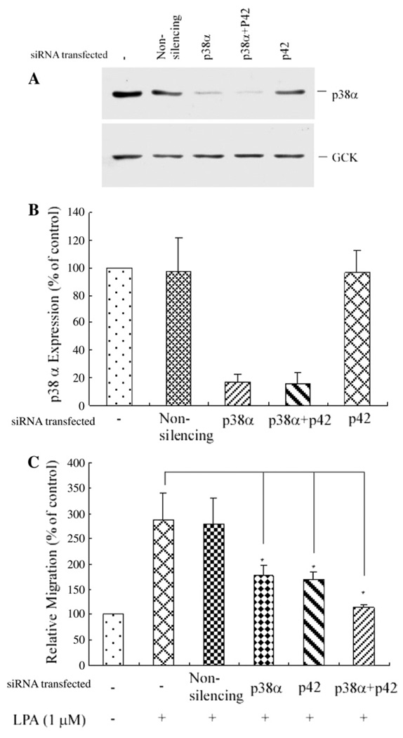
Fig. 6.
Transfection of p38α siRNA blocked endogenous p38α expression and also significantly blocked LPA-induced PC3 cell migration, and transfection of both p38α siRNA and p42 siRNA nearly completely blocked PC3 cell migration. (A) Western blot analysis of the depletion of p38α MAPK expression. Transfection of p38α siRNA alone, or transfection of p38α siRNA and p42 MAPK siRNA depleted the expression of the endogenous p38α compared with either transfection of the non-silencing siRNA (control) or transfection of p42 siRNA alone. GCK was used as a loading control. (B) Bar graph of depletion of endogenous p38α protein expression by transfection of p38α siRNA, and p38α siRNA with p42 siRNA. Data shown are from three experiments. (C) Cell migration assay. Cells were transfected with non-silencing siRNA, p38α siRNA, p42 siRNA, or p38α siRNA plus p42 siRNA. Data were mean±SE of three experiments. *p<0.05 versus LPA alone.

