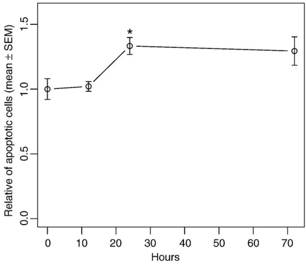
Fig. 2.
Ratio of apoptotic cells in cultured aortic rings. The thoracic aorta was surgically isolated, cultured in serum-free medium for 0–72 h, and fixed. Apoptotic cells were visualized using TUNEL staining. The percentage of apoptotic cells was evaluated by counting the stained cells/total cells. The number of apoptotic cells in each group was normalized to the 0-h group. Values are means±SEM for 4 random fields. *Denotes significant difference from the preceding time point (P<0.05).

