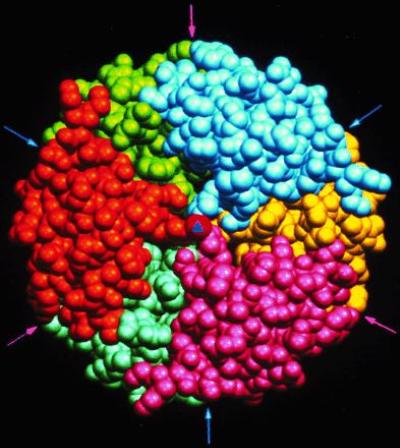
Figure 2.

The structure of the zinc insulin hexamer as defined by Hodgkin and coworkers (14). The hexamer is viewed down the exact 3-fold axis (triangle at the center); the arrows indicate positions of approximate 2-fold axes relating pairs of protomers. Each protomer is represented in a specific color, and the zinc at the center is shown in red.
