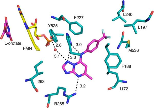
Figure 5.

Binding site of 2 bound to PfDHODH. A limited set of residues within the 4Å shell of the bound inhibitor are displayed. 2 and orotate are displayed in pink and FMN is displayed in yellow. Hydrogen bonding distances are displayed in Å. The structure (3I6R.pdb) is displayed in PyMol.
