
Figure 2.
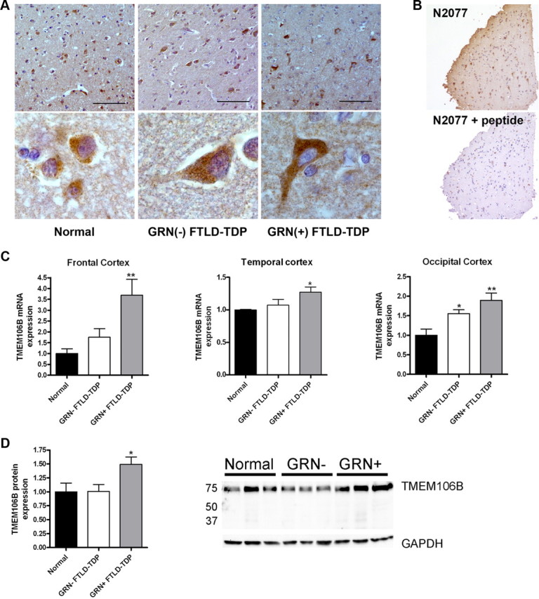
TMEM106B expression is increased in FTLD-TDP. A, Immunohistochemical staining was performed with N2077 anti-TMEM106B antibody on frontal cortex brain sections from age-matched controls (Normal), GRN(−) FTLD-TDP, and GRN(+) FTLD-TDP). GRN(+) FTLD-TDP patients had more diffuse TMEM106B staining, extending throughout the cell body and into neuronal processes. Representative lower-magnification images (top) and higher-magnification images of typical neurons from the same field (bottom) are shown. Scale bar, 100 μm. B, Neuronal staining (top) was abolished with preabsorption of N2077 with the immunizing peptide (bottom). Scale bar, 200 μm. C, Total mRNA was isolated from neurologically normal controls (n = 6), GRN(−) FTLD-TDP (n = 7), and GRN(+) FTLD-TDP (n = 5), and TMEM106B transcript expression was measured by qRT-PCR in multiple brain regions. Compared with both normal controls and to GRN(−) FTLD-TDP, GRN(+) FTLD-TDP had significantly higher levels of TMEM106B expression. Means ± SEM are shown. *p < 0.05, **p < 0.01. D, Frontal cortex protein was RIPA extracted. Equal amounts of total protein from neurologically normal controls (n = 4), GRN(−) FTLD-TDP (n = 3), and GRN(+) FTLD-TDP (n = 4) were loaded, and immunoblots were probed for TMEM106B. Corroborating our mRNA findings, GRN(+) FTLD-TDP brain showed higher levels of TMEM106B protein expression. Quantification (mean ± SEM) includes all available samples; representative subset immunoblot is also shown.