Figure 6.

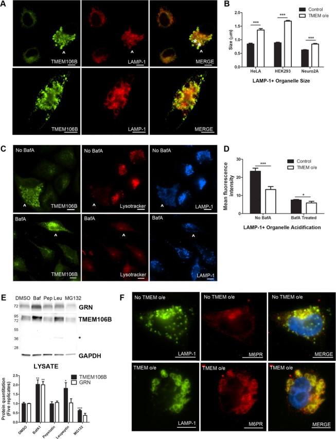
Overexpression of TMEM106B results in abnormalities in the endosomal–lysosomal pathway. A, In HeLA cells overexpressing TMEM106B (arrowhead), LAMP-1+ organelles demonstrate a general increase in size, compared with neighboring cells not overexpressing TMEM106B. In addition, overexpression of TMEM106B also results in occasional formation of large vacuolar structures ∼5 μm in diameter (asterisks indicate two vacuolar structures in top panel, also pictured throughout the cytoplasm of cell in bottom panel). While these large vacuolar structures occur only occasionally with TMEM106B overexpression (the more typical finding is enlarged ∼1.5 μm LAMP-1+ organelles), they are not seen in the absence of TMEM106B overexpression. B, Similar results were obtained in HeLAs, HEK293 cells, and in the neuronal cell line Neuro2A. Size quantitation (mean ± SEM) was performed by measuring LAMP-1+ organelle diameter on >10 40× fields containing a mixture of cells with and without TMEM106B overexpression. Because the large vacuolar structures are only occasionally seen, they were not included in the quantitation. C, HeLA cells overexpressing TMEM106B (arrowhead) showed less intense staining with Lysotracker, a dye which demonstrates greater fluorescence at lower pH, than neighboring cells not overexpressing TMEM106B (top). This effect was abrogated by treatment of cells with bafilomycin A1, an inhibitor of the vacuolar ATPase, which resulted in diminished Lysotracker fluorescence for all cells (bottom). D, Quantitation of mean fluorescence intensity for cells overexpressing TMEM106B demonstrated that Lysotracker staining was significantly less intense than in neighboring cells with normal levels of TMEM106B expression. Quantitation (mean ± SEM) was performed on >10 40× fields containing a mixture of cells with and without TMEM106B overexpression. E, Immunoblot analysis of HeLA cells treated with the vacuolar ATPase inhibitor bafilomycin A1 (Baf) showed increased intracellular levels of TMEM106B and progranulin. Treatment with the lysosomal protease inhibitor leupeptin (Leu) increased levels of TMEM106B but did not affect levels of progranulin. Treatment with the lysosomal protease inhibitor pepstatin A (Pep) did not affect either protein, while treatment with the proteasome inhibitor MG132 decreased TMEM106B levels. Representative immunoblot (top) and quantitation of five replicate immunoblots (mean ± SEM, bottom) are shown. Asterisk indicates TMEM106B 40 kDa band only seen with leupeptin treatment. F, Under normal conditions, the cation-independent M6PR does not colocalize with LAMP-1. In cells overexpressing TMEM106B, M6PR colocalizes with LAMP-1 at the limiting membrane of enlarged LAMP-1+ organelles. *p < 0.05, **p < 0.01, ***p < 0.001. For all immunofluorescence panels, TMEM106B staining was performed with N2077. Scale bar, 10 μm.
