Figure 7.

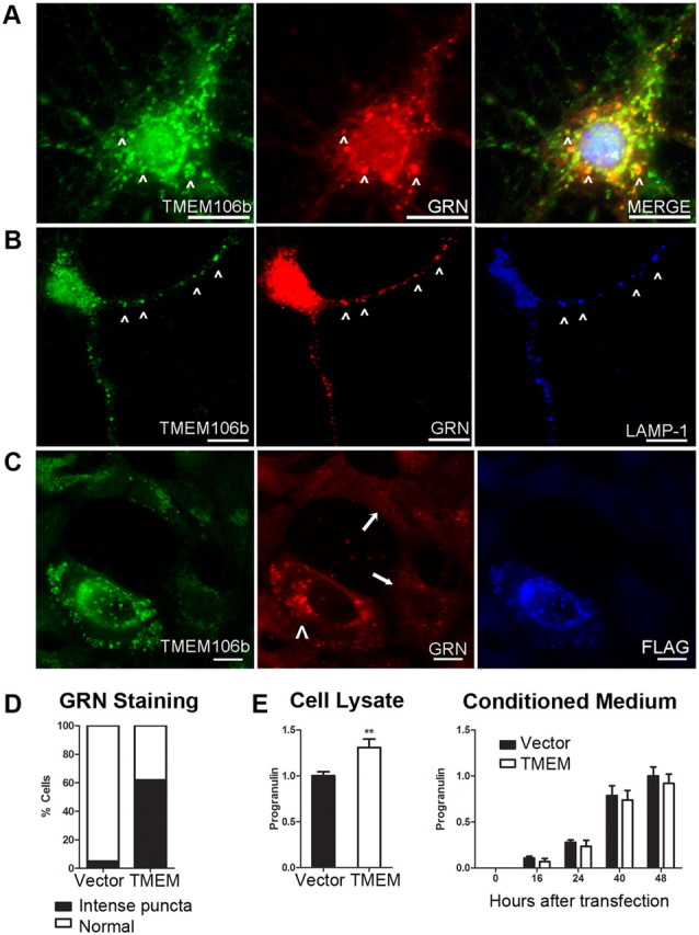
Overexpression of TMEM106B alters the compartmentalization of progranulin. A–C, Immunofluorescence microscopy performed on cells stained for TMEM106B (N2077 antibody) and progranulin. Scale bar, 10 μm. A, B, Endogenous TMEM106B (green) in nontransfected primary murine cortical neurons colocalized with progranulin (GRN, red) in the cell body (A, arrowheads), and in processes (B, arrowheads). TMEM106B and GRN colocalized within late endosomes or lysosomes, as indicated by LAMP-1 staining (B, blue). C, Progranulin (GRN, red) appearance changed under conditions of TMEM106B overexpression. Progranulin formed intensely stained cytoplasmic puncta variably colocalizing with TMEM106B (green) only in HEK293 cells overexpressing FLAG-tagged TMEM106B (arrowhead). In the absence of TMEM106B overexpression, progranulin staining was much less intense (arrows). D, More than 60% of cells overexpressing TMEM106B showed intense cytoplasmic puncta of progranulin, compared with <5% of cells with normal levels of TMEM106B expression. Assessment of progranulin staining pattern performed on six 20× fields of HEK293 cells containing a mixture of cells with and without TMEM106B overexpression. E, Intracellular (left) and extracellular/secreted (right) pools of progranulin were measured by ELISA under conditions of TMEM106B overexpression (TMEM, white bars) versus vector transfection in HEK293 cells. Progranulin measurements (mean ± SEM for five experiments) were normalized to total protein in the cell lysate, to account for differential rates of cell growth. Overexpression of TMEM106B resulted in a 30% increase in intracellular progranulin, with a trend toward decreased extracellular progranulin. Intracellular progranulin is shown measured at 48 h after transfection of TMEM106B. Extracellular progranulin is shown at baseline and indicated time periods after transfection of TMEM106B. **p < 0.01.
