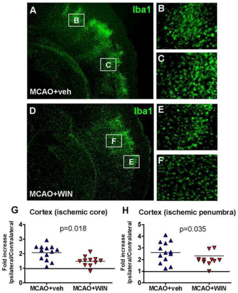
Figure 2. WIN reduces the accumulation of Iba1+ microglia/macrophages in the injured cortex after neonatal MCAO.

A–F: Immunofluorescence showing a reduced accumulation of Iba1+ cells in the ischemic penumbra (B, E) and ischemic core (C, F) in WIN-treated animals 72 hours after neonatal stroke (magnification 2.5x in A and D, 20x in B, C, E and F). G, H: Analysis of cortical Iba1 density in the ischemic core (G) and the ischemic penumbra (H) 72 hours after neonatal stroke. Data are expressed as fold increase vs the corresponding areas in the contralateral hemishere. n=11–13 per group; Student’s t-test p<0.05.
