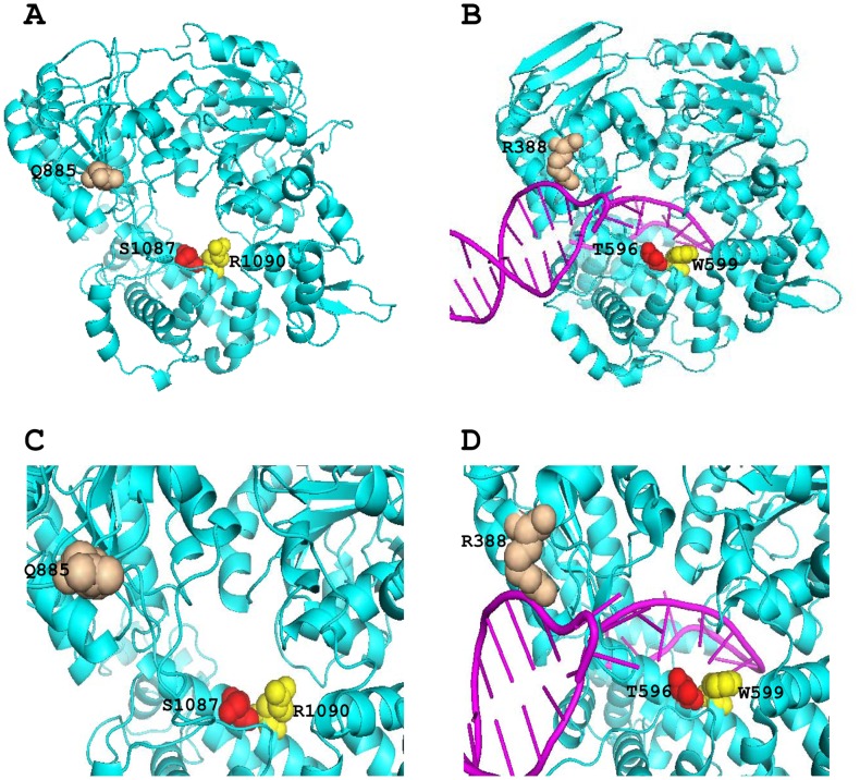
Figure 5. Structure modeling of hBrr2(477–1174).
(A) Orthogonal ribbon plots of the model of hBrr2(477–1174). Three conserved residues (Q885, S1087 and R1090) that may interact with RNA base are shown as space-filling spheres, and colored as wheat, red and yellow respectively. (B) Orthogonal ribbon plots of the Hel308 DNA helicase [2] (PDB ID: 2p6r). Cyan, Hel308; pink, DNA. Three DNA interacting residues R388, T596 and W599 in Hel308 which correlate to Q885, S1087 and R1090 in human respectively are shown as space-filling spheres, and colored in order as wheat, red and yellow. (C) A close view of the central pore in the model, highlighting the three conserved residues (Q885, S1087 and R1090). (D) A close view of the path of DNA strand through the central pore, highlighting the three related residues (R388, T596 and W599) implicated in DNA binding.

