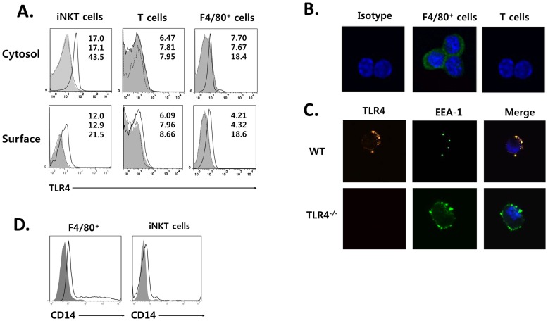
Figure 1. iNKT cells constitutively express TLR4 on the cell surface and in the endosomal compartment.
(A) TLR4 expression was analyzed on gated α-GalCer/CD1d tetramer-CD3+ T cells, α-GalCer/CD1d tetramer+ iNKT cells, and F4/80+ macrophages from B6 (solid line) or TLR4−/− mice (gray) compared with an isotype-matched control IgG (dotted line) by flow cytometric analysis. Numbers in diagrams represent mean fluorescence intensity (top for control, middle for TLR4−/− mice, bottom for B6 mice). (B) Sorted iNKT cells and F4/80+ macrophages were stained with anti-TLR4 mAb (green) or isotype-matched control IgG, and DAPI (blue) (C) Sorted iNKT cells were stained with anti-TLR4 mAb or isotype-matched control IgG (red), and EEA-1 (early endosome marker; green) and DAPI (blue). (D) CD14 expression was analyzed on gated α-GalCer/CD1d tetramer+ iNKT cells and F4/80+ macrophages from B6 mice (solid line) as compared with an isotype-matched IgG control (gray). Data are representative of three independent experiments.

