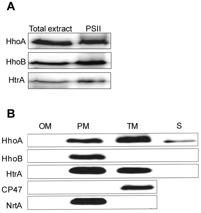
Figure 2. Subcellular localization of the Synechocystis Deg proteases.
(A) Total wild-type cell extract and a PSII-enriched fraction isolated from the HT3 strain of Synechocystis 6803 were analyzed by SDS-PAGE, and by immunostaining using antibodies directed against recombinant HhoA, HhoB, or HtrA. Of the total cell extract, samples corresponding to 0.1 µg of chlorophyll were loaded for immunostaining with anti-HhoA and samples corresponding to 0.5 µg were loaded for anti-HhoB and anti-HtrA. Of the PSII-enriched fraction, samples corresponding to 5 µg of chlorophyll were loaded in each lane. (B) Outer membrane (OM), plasma membrane (PM), thylakoid membrane (TM), and soluble protein fraction (S) were isolated from Synechocystis 6803 by gradient centrifugation and two-phase partitioning, separated by SDS-PAGE, and immunostained using antibodies directed against recombinant HhoA, HhoB, or HtrA. Purity of the membranes was determined by using antibodies directed against the PSII protein CP47 and against NrtA, a component of the nitrate transporter; 15 µg of protein was loaded in each lane.

