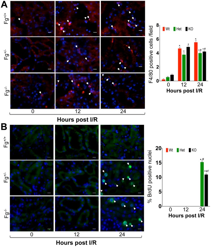
Figure 3. Heterozygous and knockout Fg mice exhibit efficient immune cell clearance coupled with robust tubular epithelial cell proliferation.
Fixed frozen sections following IRI were stained for A) macrophage F4/80 (red). Number of F4/80 cells per 60X field is represented graphically on the right of the photomicrograph. B) BrdU positive cells (green) by immunofluorescence. Percentage of positive staining for BrdU positive nuclei is represented graphically on the right of photomicrographs. Arrowheads indicate positive cells/nucleus and bar represents 10 µm. *represents p<0.05 in comparison to sham; #represents p<0.05 as compared to wild type within the time point and !represents p<0.05 as compared to heterozygous within the time point as determined by one-way ANOVA. Bar represents 10 µm.

