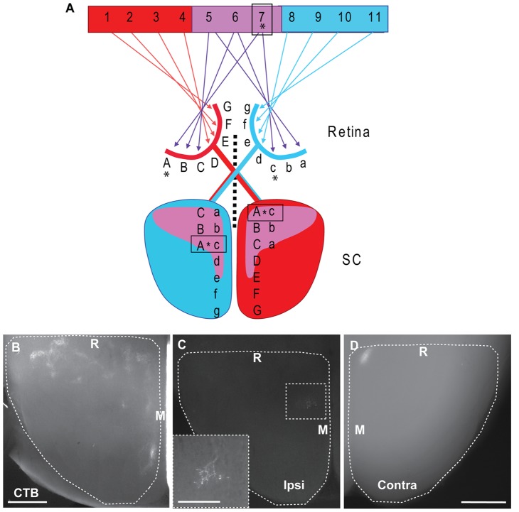
Figure 1. Schematic diagram and DiI tracing illustrating the visuotopic organisation of the retinocollicular pathway from ventrotemporal retina in the mature mouse.
A: Left eye (red) G-A, representing visual field 1–7 projects topographically to the contralateral SC (red). Similarly, the right eye (blue) a–g, represents visual field 5–11 which projects topographically to the left SC (blue). The binocular field (centre, purple) 5–7 is represented by the ventrotemporal retina of both eyes (C-A, red & a–c, blue) and is the origin of the ipsilateral projection. The ipsilateral projection maps in reverse retinotopic order to produce an aligned map in the SC. Note that a given point in the visual field (e.g. 7,*) falls on different locations in the two retinas (red A & blue c, *). In order to generate an aligned map, these different retinotopic locations must project to the same region in the target (boxed regions, *). Conversely, the same retinotopic locations (red A & blue a) map to two different locations in the SC. When considering the ipsilateral and contralateral projections arising from a given retinotopic location from a single eye (e.g. red A, *), these will project to distinct regions in the contralateral and ipsilateral SC. Note the differing positions of the representation of red A in each collicular hemisphere (boxes, *). B: Horizontal section, approximately 300 µm below the surface of a flattened SC, from a P28 mouse following an injection of CTB into the ipsilateral eye to label all RGC axons. Ipsilateral terminals are arranged in clusters in the rostromedial region of the SC. The majority of the ipsilateral projection is visible in this single section. C–D: Horizontal section through the ipsilateral (ipsi) SC (C) and a whole-mount preparation of the contralateral (contra) SC (D) following a focal injection of DiI into the VTC from a P28 mouse. A dense contralateral TZ is visible at the rostromedial corner of the SC in D. In the ipsilateral SC (C) the TZ is offset caudolaterally from the contralateral TZ. The ipsilateral TZ is much less dense than the corresponding contralateral TZ and is shown at higher power in the inset. Scale bars: 500 µm. Scale in D also applies to C. Scale in inset in B: 200 µm. R: Rostral, M: Medial. Dashed lines delineate the borders of the SC.

