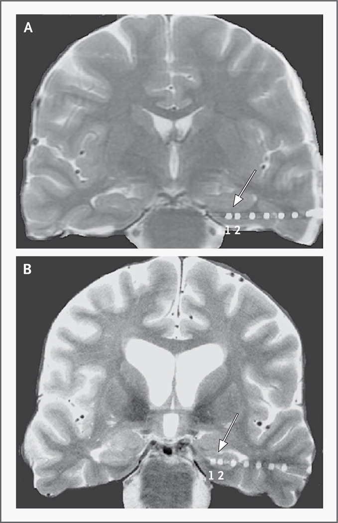
Figure 1. High-Resolution Magnetic Resonance Imaging (MRI) in Two Subjects with Implanted Electrodes.
Coronal high-resolution MRI scans show electrodes imaged on postoperative computed tomographic (CT) scans coregistered to the MRI scans. The two most distal electrodes (numbered 1 and 2, with 1 marking the most distal electrode) are shown in the left entorhinal region in one subject (Panel A) and the ipsilateral hippocampus in another subject (Panel B). For all subjects, the two most distal electrodes were used for stimulation.

